- Issues
- About the Journal
- News
- Cooperation
- Contact Info
Issue. Articles
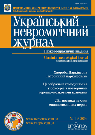
№1(38) // 2016

1. Reviews
|
Notice: Undefined index: picture in /home/vitapol/ukrneuroj.vitapol.com.ua/en/svizhij_nomer.php on line 74 Notice: Undefined index: pict in /home/vitapol/ukrneuroj.vitapol.com.ua/en/svizhij_nomer.php on line 75 Perinatal hypoxic-ischemic encephalopathy and experimental approaches to its correctionK. V. YatsenkoBogomoletz Institute of Physiology NAS Ukraine, Кyiv |
|---|
Keywords: hypoxic-ischemic encephalopathy, neuroprotection.
Notice: Undefined variable: lang_long in /home/vitapol/ukrneuroj.vitapol.com.ua/en/svizhij_nomer.php on line 188
2. Lectures
|
Notice: Undefined index: picture in /home/vitapol/ukrneuroj.vitapol.com.ua/en/svizhij_nomer.php on line 74 Notice: Undefined index: pict in /home/vitapol/ukrneuroj.vitapol.com.ua/en/svizhij_nomer.php on line 75 Modern views on etiopathogenesis, diagnosis and treatment of Parkinson’s disease and secondary parkisonismO. A. MialovytskaO. O. Bogomolets National Medical University, Kyiv |
|---|
Keywords: Parkinson’s disease, syndrome of parkinsonism, exstrapyramidal system, diagnosis, treatment.
Notice: Undefined variable: lang_long in /home/vitapol/ukrneuroj.vitapol.com.ua/en/svizhij_nomer.php on line 188
3. Original researches
|
Notice: Undefined index: picture in /home/vitapol/ukrneuroj.vitapol.com.ua/en/svizhij_nomer.php on line 74 Notice: Undefined index: pict in /home/vitapol/ukrneuroj.vitapol.com.ua/en/svizhij_nomer.php on line 75 ApoE gene polymorphism, lipid profile and psychodiagnostic status in patients with dyscirculatory encephalopathyN. V. Lytvynenko, O. Ye. PalyenkaUkrainian Medical Stomatological Academy, Poltava |
|---|
Methods and subjects. A comprehensive clinical, psychodiagnostic, biochemical and genetic study of 80 patients with dyscirculatory encephalopathy was performed during 2013 — 2014 at Poltava region hospital named after N. V. Sklifosovskiy.
Results. Genotyping of polymorphism of ApoE gene revealed the most common genotype 3ε3/ε2 (58 patients (72.5 %)). ε4/ε3 genotype was registered in 12 patients (15 %), genotype ε3/ε3 6 people (7, 5 %). When comparing the demographic variables in groups of patients with the genotype ε3/ε3 andε3/ε2 the female prevalence was revealed, ε4/ε3 genotype was mostly determined in males. Patients of all groups had no significant age differences. In all three groups patients had a BMI greater than 30 kg/m2, which indicates the presence of obesity. At the ε3/ε3 genotype patients demonstrated the highest rate. The maximum total cholesterol and triglycerides rate in blood was in patients with ε4/ε3 genotype.
Conclusions. Genotype ε3/ε2 (72.5 %) was presented at the predominant number of patients. Genotype ε3/ε3 and ε4/ε3 demonstrated high rate of BMI, waist volume, hips and waist volume correlation comparing with genotype ε3/ε2. The maximum total cholesterol and triglycerides rate in blood was in patients with ε4/ε3 genotype . In accordance with MMSE test patients of all groups suffered from mild cognitive impairments. Evidenced decrease in the short and long memory as revealed in genotype ε3/ε2 patients, elevated level of personal anxiety was observed in genotype ε3/ε3 patients.
Keywords: apolipoprotein E, gene apolipoprotein E, allele ε2, ε3, ε4, polymorphism, lipid profile, cognitive impairments.
Notice: Undefined variable: lang_long in /home/vitapol/ukrneuroj.vitapol.com.ua/en/svizhij_nomer.php on line 188
4. Original researches
|
Notice: Undefined index: picture in /home/vitapol/ukrneuroj.vitapol.com.ua/en/svizhij_nomer.php on line 74 Notice: Undefined index: pict in /home/vitapol/ukrneuroj.vitapol.com.ua/en/svizhij_nomer.php on line 75 Clinical and diagnostic aspects of cognitive disorders in patients with atrioventricular blockadeS. М. StadnikMilitary Clinical Medical Centre of the Western Regions, Lviv |
|---|
Methods and subjects. Examined 107 patients (mean age 63.2 ± 10.8 years) with AV blockade varying degrees. A comparative analysis of neuropsychological characteristics and lipid profile in patients was studied.
Results. The frequency of cognitive disorders in patients with AV blockade I was 27 (52.9 %), including: mild— 18 (66.7 %) and moderate — 9 (33.3 %). In patients with AV blockade II cognitive disorders were detected in 24 (68.6 %) patients, including mild —12 (34.3 %), moderate in 7 (20.0 %), severe—in 5 (11.4 %) of patients. In patients with AV blockade III cognitive disorders were detected in 19 (90.5 %) patients, including mild — 3 (14.3 %), moderate — in 13 (61.9 %) of the severe — 5 (23.8 %) patients. In the quantitative analysis of cognitive functions scale MMSE all patients with AV blockade II and III were statistically significantly different from patients with AV blockade I according to the results of the sub tests «account», «memory», «the repetition of the phrases», «praxis». It has been shown statistically significant increases in total cholesterol, cholesterol of low¬density lipoprotein and triglycerides in patients with AV blockade II and III compared with patients with AV blockade I.
Conclusions.The frequency of cognitive disorders in patients with AV blockade increased with progression of the disease. Risk factors are old age, II and III degree of the disease, high cholesterol low density lipoprotein. Adverse was a combination of several risk factors for cognitive problems in patients with AV blockade. Increased severity of cognitive disturbance occurs in patients not adhering to treatment and control heart rate.
Keywords: cognitive disorders, atrioventricular blockade, blood lipid profile.
Notice: Undefined variable: lang_long in /home/vitapol/ukrneuroj.vitapol.com.ua/en/svizhij_nomer.php on line 188
5. Original researches
|
Notice: Undefined index: picture in /home/vitapol/ukrneuroj.vitapol.com.ua/en/svizhij_nomer.php on line 74 Notice: Undefined index: pict in /home/vitapol/ukrneuroj.vitapol.com.ua/en/svizhij_nomer.php on line 75 Cerebrovascular reactivity in the initial manifestations of chronic cerebral ischemiaМ. А. TrishchinskaP. L. Shupyk National Medical Academy оf Postgraduate Education of Health Ministry of Ukraine, Kyiv |
|---|
Methods and subjects. We have examined 172 individuals (44 men and 128 women), aged 50.0 ± 0.45 with initial manifestations of the chronic cerebral ischemia. Patients were divided into two groups depending on neuro visual changes of the brain of vascular origin, and patients of group 2 were divided into two (A and B) groups with the presence of cerebral atrophy. All patients underwent general clinical, clinical-neurological, instrumental and clinical laboratory tests. To study cerebrovascular reactivity we performed transracial duрplex scanning using multidirectional metabolic and myogenic functional tests.
Results. Patients of different groups (p < 0.001) were statistically differed by index reactivity (ІR) obtained in the metabolic and myogenic multidirectional tests in carotid pool. By increasing the degree of brain damage at initial manifestations chronic cerebral ischemia, IR in response to metabolic and myogenic stimuli decreased too within autoregulation range. The reaction to vazodilatatory tests was minor than the reaction in response to vasoconstrictor stimuli.The variability coefficient of parameters in response to both vasodilator and vasoconstrictor stimuli increased with the development of chronic brain ischemia towards unity, decreasing the variability of cerebral reactivity.
Conclusions. Indications of cerebrovascular reactivity in patients with initial manifestations of chronic brain ischemia have clinical and diagnostic value, even within autoregulation range. Therefore with IR approaching to one or another range we are able to determine the degree of compensatory mechanisms stress aimed at supporting homeostasis of cerebral blood flow.
Keywords: cerebrovascular reactivity, the initial signs, chronic cerebral ischemia.
List of references:
1. Лелюк В. Г., Лелюк С. Э. Методика ультразвукового исследования сосудистой системы: технология сканирования, нормативные показатели: Метод. рекомендации. — М., 2002. — С. 40.
2. Лелюк В. Г., Лелюк С. Э. Церебральное кровообращение и артериальное давление. — М.: Реальное время, 2004. — С. 304.
3. Лелюк С. Э., Лелюк В. Г. Методические аспекты ультразвукового исследования цереброваскулярной реактивности в норме и при атеросклеротическом поражении брахеоцефальных артерий: Учеб. пособие. — М.: РМАПО, 2009. — С. 28.
4. Москаленко Ю. Е., Бекетов А. И., Орлов Р. С. Мозговое кровообращение. — Л.: Наука, 1988.
5. Muller M., Voges M., Piepgras U. Assessment of cerebral vasomotor reactivity by transcranial Doppler ultrasoundand breath-holding. A comparison with acetazolamide as vasodilatory stimulus // Stroke. — 1995. — Vol. 26. — P. 96 — 100.
6. Wardlaw J., Smith E., Cordonnier Ch. et al. Neuroimaging standards for research into small vessel disease and its contribution to ageing and neurodegeneration // Lancet Neurol. — 2013. — N 12. — P. 822 — 838.
Notice: Undefined variable: lang_long in /home/vitapol/ukrneuroj.vitapol.com.ua/en/svizhij_nomer.php on line 188
6. Original researches
|
Notice: Undefined index: picture in /home/vitapol/ukrneuroj.vitapol.com.ua/en/svizhij_nomer.php on line 74 Notice: Undefined index: pict in /home/vitapol/ukrneuroj.vitapol.com.ua/en/svizhij_nomer.php on line 75 Influence of pro- and antioxidative system status on the vascular and platelet hemostasis in patients in the early recovery period of ischemic strokeV. R. GerasymchukIvano-Frankivsk National Medical University |
|---|
Methods and subjects. The study involved 52 patients 1 month following atherothrombotic hemispheric IS. Status of vascular and platelet hemostasis was assessed by degree, rate and time of platelets aggregation, number of platelets and activity of von Willebrand factor (vWF). Status of pro- and antioxidative system was evaluated by the content oxidative modification of proteins (OMP) products, the activity of glutathione peroxidase (GP) and glutathione reductase (GR).
Results. In patients after stroke significant elevated level of OMP products was observed against the background of decreased activity of GP and GR (p < 0.05). Increased activity of oxidation processes with high concentrations of OMP products was accompanied with activation of vascular and platelet hemostasis with an increased vWF activity (r = 0.61; p = 0.009) and platelets aggregation rate (r = 0.49; p = 0.031).
Conclusions. Disbalance of pro- and antioxidative systems in the early recovery period of IS correlates with the vascular and platelet hemostasis indexes with a tendency to increase rate of platelet aggregation and von Willebrand factor activity, that can influence the course of the recovery period of IS and increase the risk of recurrent stroke.
Keywords: ischemic stroke, early recovery period, vascular and platelet hemostasis, von Willebrand factor, oxidative modification of proteins.
Notice: Undefined variable: lang_long in /home/vitapol/ukrneuroj.vitapol.com.ua/en/svizhij_nomer.php on line 188
7. Original researches
|
Notice: Undefined index: picture in /home/vitapol/ukrneuroj.vitapol.com.ua/en/svizhij_nomer.php on line 74 Notice: Undefined index: pict in /home/vitapol/ukrneuroj.vitapol.com.ua/en/svizhij_nomer.php on line 75 Structural сhanges of the brain in patients with cognitive impairments against the background of metabolic syndromeT. I. Nasonova1, A. L. Susenko21 P. L. Shupyk National Medical Academy оf Postgraduate Education of Health Ministry of Ukraine, Kyiv |
|---|
Methods and subjects. Clinical and neurological examinations were carried out for 59 patients with cerebrovascular diseases. 38 patients in the study group had evidence of metabolic syndrome (MS) and 21 patients without the MS comprised a control group. All patients underwent clinical and neurological examinations, cognitive impairment evaluation on scales MMSE, MOCA, Schulte’s tables. By means of 1.5 T MRI system MAGNETOM Avanto 1.5 T machine by Siemens SQ the following measurements were performed : the thickness of grey matter in the frontal and parietal lobes in cm right and left; the size of the lateral ventricles of the brain right and left, in mm (anterior horn, body, posterior horn), the size of the third ventricle of the brain; the presence of foci and their sizes.
Results. We identified the presence of brain atrophy signs in all patients, in particular, the reduction of the thickness of the cortex in the frontal and parietal lobes, enlargement of the ventricular system of the brain (increase in the size of the anterior horns and bodies of the ventricles). The thickness of grey matter (cortex) in the frontal lobe of patients of the main group was (median and microtile interval) 0.2675 [0.180 — 0.315] against 2.800 [0.360 — 4.210] in the control group. The thickness of the cortex in the temporal region was: in the main group 0.27050 [0.20275 — 0.35000] against control group 0.31000 [0.25400 — 0.34800]. In the frontal lobes of the thickness of the grey matter (cortex) was significantly less in patients with cerebrovascular diseases against the background of MS (p < 0.05). The significant difference (p < 0.05) was determined between sizes of the right front corner in mm (3.2550 [0.7950 — 5.7450] in the main group against 2.8000 [0.3600 — 6.2100] in controls) and body of the left lateral ventricle, in mm (5.74 [2.01 — 8.65] in the main group against 4.8 [2.2 — 7.65] in control), as well as the right lateral ventricle, in mm (5.9850 [at 1.4600 — 9.2050] in the primary against 5.0300 [0.7500 — 8.0200] in the control group). The study of cognitive functions according to the scale of MOCA (25.25 [24 — 27] demonstrated 27 [25 — 28] in the main group against in the control and MMSE (26.25 [25 — 27] in the main group and 27.5 [25 — 28] in the control). It implied significantly (p < 0.05) marked cognitive impairment in patients of middle age in the main group. The inverse ratio between indicators of the thickness of the cortex of the frontal lobe and results of cognitive function on the MOСA scale was determined. The index of correlation amounted to rs = –0.39. The module of correlation coefficient was within the limits of medium strength.
Conclusions. In patients with cerebrovascular diseases against the background of MS the statistically significant cognitive performance impairment was determined in comparison with similar indicators in patients with cerebrovascular diseases with no signs of MS. In patients with cerebrovascular diseases the signs of atrophic process of the brain were observed, but in patients with MS these symptoms were more marked. The ratio between cognitive impairment and thickness of the cortex in the frontal lobe of the brain was estimated in patients with cerebrovascular diseases against the background of MS.
Keywords: cognitive impairment, brain atrophy, metabolic syndrome.
Notice: Undefined variable: lang_long in /home/vitapol/ukrneuroj.vitapol.com.ua/en/svizhij_nomer.php on line 188
8. Original researches
|
Notice: Undefined index: picture in /home/vitapol/ukrneuroj.vitapol.com.ua/en/svizhij_nomer.php on line 74 Notice: Undefined index: pict in /home/vitapol/ukrneuroj.vitapol.com.ua/en/svizhij_nomer.php on line 75 Comparative evaluation of the background neurological deficits in patients with acute ischemic stroke against the background the primary metabolic syndrome presenceO. M. Dzyuba1, V. V. Babenko1, 21 O. O. Bogomolets National Medical University, Kyiv |
|---|
Methods and subjects. The study involved 160 patients with ischemic stroke (men — 103 (64.4 %), women — 57 (35.6 %)) aged from 39 years to 91 (mean age — 66.5 ± 9.4 years). Patients were divided into two groups. The focus group includes 102 patients (men — 68 (66.7 %), women — 34 (33.3 %), mean age — 64.4 ± 9.4 years), who had IS against the MS background. Second, the control group, consisted of 58 patients without MS (men — 35 (60.3 %), women — 23 (39.7 %), mean age — 70.5 ± 9.3 years), who experienced the cerebral infarction against the background of hypertension, atherosclerosis, coronary heart disease, atria fibrillation. A comprehensive clinical and neurological examinations, MRI and/or cerebral computer tomography were conducted. Evaluation of background neurologic deficit was carried out at the time of patient admission by NIHSS scale, functional status of patients was assessed by Barthel Index and modified Rankin scale.
Results. According to the study, it was established that MS sets the stage for the emergence of a cerebral accident, and significantly affects the intensity of the background neurological deficit, intensifying it. Patients with metabolic syndrome demonstrated the prevalence of significant lesions in the system of the middle cerebral artery through a complete heart attacks, brain stem and deep branches lesions. The infarct volume in the carotid was prevalent in patients of the focus group, intensifying neurological disorders. The significant prevalence of lacunar infarcts with vertebrobasilar lesions pool is a proof for metabolic abnormalities impact that cause the immediate occurrence of MS.
Conclusions. MS not only paves the way for the emergence of the primary form of acute ischemic stroke cerebral angiopathy vessels, but also intensifies the background neurological deficits significantly due to high volume of infarction focus.
Keywords: ischemic stroke, metabolic syndrome, background neurologic deficit.
Notice: Undefined variable: lang_long in /home/vitapol/ukrneuroj.vitapol.com.ua/en/svizhij_nomer.php on line 188
9. Original researches
|
Notice: Undefined index: picture in /home/vitapol/ukrneuroj.vitapol.com.ua/en/svizhij_nomer.php on line 74 Notice: Undefined index: pict in /home/vitapol/ukrneuroj.vitapol.com.ua/en/svizhij_nomer.php on line 75 Bioadaptive control in complex treatment of psycho-emotional impairments among patients with chronical cerebral ischemiaА. V. DemchenkoZaporizhzhya State Medical University |
|---|
Methods and subjects. 55 patients with CCI were evaluated for clinical and neuropsychological, instrumental and statistical methods of investigation. Patients were divided into 2 groups depending on the scheme of treatment: basic (n = 30) and control (n = 25). The complex treatment of patients from the basic group involved the traditional therapy combined with bioadaptive control in the form of united courses of alpha-stimulating and temperature-myograhic trainings based on biological feedback (BFB).
Results. After complex treatment of patients with CCI using BFB trainings the following data were achieved: decreased the number of complaints for headache (р = 0.0002), dizziness (р = 0.001), staggering while walking (р = 0.003), sleep disturbance (р = 0.094), irritability (р = 0.027), anxiety (p = 0.013), the concentration of attention and memory (р = 0.001) improved in comparison with control group, where the complaints for headache, dizziness and staggering while walking intensified. The level of reactive anxiety (р < 0.01) and personal anxiety (р < 0.001) was reduced significantly in accordance with Spielberger test. Depressive symptoms regressed among 40 % of patients from the basic group against 12 % of patients from the control group. After conducting BFB trainings among patients with CCI the reliable reduce of latent period of cognitive evoked potential Р300 in accordance with Fridman (р) was observed: F3 = 0.003; F4 = 0.015; Fz = 0.039; C3 = 0.008; C4 = 0.016; Cz = 0.011; P3 = 0.010; P4 = 0.017; Pz = 0.007 and the significant increase of alpha-activity in the right frontal and parietooccipital, temporal areas was determined.
Conclusions.Bioadaptive control is an efficient method of complex treatment for anxiety-depressive disorder, correction of cognitive impairments and changes of neurophysiologic rates among patients with CCI.
Keywords: chronical cerebral ischemia, bioadaptive control, psycho-emotional sate, cognitive impairments.
Notice: Undefined variable: lang_long in /home/vitapol/ukrneuroj.vitapol.com.ua/en/svizhij_nomer.php on line 188
10. Original researches
|
Notice: Undefined index: picture in /home/vitapol/ukrneuroj.vitapol.com.ua/en/svizhij_nomer.php on line 74 Notice: Undefined index: pict in /home/vitapol/ukrneuroj.vitapol.com.ua/en/svizhij_nomer.php on line 75 Features of cerebral hemodynamics in boxers with repeated traumatic brain injuriesА. V. Muravskiy1, M. V. Globa2, G. V. Michal21 P. L. Shupyk National Medical Academy оf Postgraduate Education of Health Ministry of Ukraine, Kyiv |
|---|
Methods and subjects. Ultrasound tests of head and neck vessels was conducted for 156 boxers aged 17 to 42 years, who had history of repeated mild TBI. 30 healthy people from the control group of similar age were examined. Patients were analyzed according to gender, weight category, the number of matches.
Results. For boxers it is typical to have the increased diameter of extracranial vessels, the incidence of deformation of the vertebral artery (V2 segment), venous disorders, vasospasm. The focus group was characterized by the increase of blood flow velocity in the extracranial departments carotid and decrease on vertebrobasilar (VBB) pool with the change of the index of peripheral resistance, in the intracranial departments carotid was typical of the reduction of blood flow velocity without changing the index of peripheral resistance. Most boxers demonstrated signs of venous circulatory distress in the form of a deviation of the speed indicators in the internal jugular vein and the veins of Rosenthal.
Conclusions. Boxers who had history of repeated mild TBI, suffer from hemodynamic disturbances. The running deformation, increased diameter of extracranial vessels, changes in blood flow through the vessels of the carotid and VBB pools, deviations of indicators of vascular tone, venous disorders are typical for boxers.
Keywords: traumatic brain injury, boxer, ultrasound of the blood vessels of the head and neck.
Notice: Undefined variable: lang_long in /home/vitapol/ukrneuroj.vitapol.com.ua/en/svizhij_nomer.php on line 188
11. Original researches
|
Notice: Undefined index: picture in /home/vitapol/ukrneuroj.vitapol.com.ua/en/svizhij_nomer.php on line 74 Notice: Undefined index: pict in /home/vitapol/ukrneuroj.vitapol.com.ua/en/svizhij_nomer.php on line 75 Diagnosis of the spinal roots and cervical nerves tumorsE. I. Slynko1, Yu. V. Derkach1, O. M. Кhonda21 SI «Institute of Neurosurgery named after acad. A. P. Romodanov of NAMS of Ukraine», Kyiv |
|---|
Methods and subjects. The study included 60 patients with tumors of spinal nerves of the cervical spinal cord that were treated in Institute of Neurosurgery named after acad. A. P. Romodanov of NAMS of Ukraine by 2006 — 2016. Their medical history, clinical data were analyzed. We have singled out radicular sensory impairments and radicular motor segmental infringement. Over time, some patients experienced accompanying sensory and motor violations. In our study, we carried out an assessment of patients for symptoms of pain, segmental sensory and motor disorders; sensitive and motor conduction disorders. Assessment of tumor location, their size, features a compression of the spinal cord or its roots, and location in relation to the bony structures of the spine was performed by means of MRI.
Results. Among 60 patients with spinal tumors, brain nerve cervical spinal neurinoma were found in 44 patients, 7 patients had neurofibromas, peryneuromas — 2, malignant tumors of peripheral nerves — 3, parahanhliomy — 2, neyrosarcoma — 1, hanglioblastoma — 1. According to our data, neuromas growth rate reached 2.4 mm for 1 year (fluctuations 1.8 — 3 mm) and it was relatively stable. Neurofibromas growth rate ranged from 1.8 mm to 37 mm by 1 year, accounting in average 16 mm per year, and it was not stable. With over their existence, they changed their growth rate. The rate of growth of other tumors was significantly faster, unstable; but due to small number of tumors it was not possible to evaluate. The maximum size of tumors in the vertebral canal on MRI axial slices ranged from 2.5 to 21 mm, in average 8.2 mm. Tumors sometimes caused channel expansion, but it was insignificant. However, the size of the tumor, “on over” ranged from 3 mm to 379 mm, in one case reaching the level C1 to C7 vertebra. Fluctuations in the size of intervertebral foramen tumors was from 1 mm to 38 mm, in average 15 mm. Paravertebral tumor volume ranged from 2.4 mm3 to 8984 mm3, accounting in average of 892 mm3.
Conclusions. Early diagnosis of spinal nerves tumors is very important for further treatment. Clinically, in all cases, spinal tumors originated from segmental disorders. While growing tumor in the spinal cord, motor violations may accompany segmental disorders. The main reason for diagnostic errors is the onset tumor segmental disorders, complicating differential diagnosis with compression symptoms of degenerative processes of the spine. Timely evaluation of segment and conduction impairments, which correlate with MRI data, provides with the opportunity to perform early diagnostics of spinal nerves tumors in order to determine the surgery methods.
Keywords: tumors of the spinal nerves, neurological manifestations, MRI diagnosis.
Notice: Undefined variable: lang_long in /home/vitapol/ukrneuroj.vitapol.com.ua/en/svizhij_nomer.php on line 188
12. Original researches
|
Notice: Undefined index: picture in /home/vitapol/ukrneuroj.vitapol.com.ua/en/svizhij_nomer.php on line 74 Notice: Undefined index: pict in /home/vitapol/ukrneuroj.vitapol.com.ua/en/svizhij_nomer.php on line 75 «Cerebral form» of the lung cancerYu. V. Dumanskiy1, O. Yu. Stolyarova2, O. V. SynІachenko1, E. D. Iegudina3, V. A. Stepko11 Donetsk National Medical University of Maxim Gorky, Lyman |
|---|
Methods and subjects.The study included 1071 patients with lung cancer at the age of 24 to 86 years. None of the examined patients has been operated previously on account of the underlying disease, all patients received radiation therapy after establishment of the diagnosis, 3/4 of them received combined radio chemotherapy.
Results. Вrain metastases (BM) occurs in 8 % of patients with LC (in 2.2 times more common in women), that is affected by the peripheral form, histological variant (squamous cell and large cell carcinoma) and integrated gravity of the tumour process, the presence of exudative chancrous pleurisy, the germination of the tumour in to the mediastinum and concomitant diabetes mellitus type 2. The severity of BM in lung cancer directly correlates with metastasis in the supraclavicular, in guinal and retroperitoneal lymph nodes, pericardium, adrenal glands, abdominal wall, liver and pancreas. The predictor of BM in patients with lung cancer may be elevated parameters of the transforming growth factor β1 in the blood, vascular endothelial growth factor, and osteopontin. In 6 % of complications’ cases stroke of different severity was observed due to the radio chemotherapy, which is closely associated with hypertension and diabetes, the form of LC, the number of metastases in the lymph nodes (but not in the brain), and with using of antitumor alkylating agents in treatment.
Conclusions. The socalled «brain form» of LC is characterized by greater degree of severity of the disease, requires correction of drug chemotherapy, determines the survival of the patients, that is less in patients with BM.
Keywords: cancer, lung, brain, metastases.
Notice: Undefined variable: lang_long in /home/vitapol/ukrneuroj.vitapol.com.ua/en/svizhij_nomer.php on line 188
13. Original researches
|
Notice: Undefined index: picture in /home/vitapol/ukrneuroj.vitapol.com.ua/en/svizhij_nomer.php on line 74 Notice: Undefined index: pict in /home/vitapol/ukrneuroj.vitapol.com.ua/en/svizhij_nomer.php on line 75 Benign multiple sclerosis: criteria and course peculiaritiesO. D. ShulgaVolyn Regional Clinical Hospital, Lutsk |
|---|
Methods and subjects. 80 patients met the criteria for benign MS. Women prevailed — 53 (66.25 %). The women: men ratio was 1.96 : 1. According to the severity by EDSS scale, patients were divided into three groups: the first group included patients with a degree of EDSS ≤ 2.0 points (n = 16), the second group — 2.5 — 3.5 points (n = 41), and the third group — 4.0 points (n = 23). Groups were comparable in age and sex. All patients had a disease duration of more than 10 years and remained partially or fully employed.
Results. It was found that the second group of patients had the higher score by the pyramid (p < 0.001), cerebellar (p < 0.001) functional scales and EDSS score (p < 0.001). The patients of the third group experienced a statistically higher score for brainstem (p < 0.05), bowel and bladder (p < 0.01), cerebral (p < 0.05) functional scales and ambulation (p < 0.05) as well as the total score for EDSS (p < 0.001) compared with the first and second groups.
Conclusions. It was established that the incidence of benign MS according to the Registry was 12.59 %. In patients with benign MS with EDSS score less than 3.0, the involvement of cerebellar and pyramidal functional systems are dominant. Brainstem and cerebral function as well as bowel and bladder are dominant in patients with EDSS score more than 3.0 points. Thus, the absence of marked motor and cerebellar deficit in patients with benign MS with preserved mobility does not always reflect the true extent of disability.
Keywords: benign multiple sclerosis, criteria, motor and non-motor symptoms.
Notice: Undefined variable: lang_long in /home/vitapol/ukrneuroj.vitapol.com.ua/en/svizhij_nomer.php on line 188
14. Original researches
|
Notice: Undefined index: picture in /home/vitapol/ukrneuroj.vitapol.com.ua/en/svizhij_nomer.php on line 74 Notice: Undefined index: pict in /home/vitapol/ukrneuroj.vitapol.com.ua/en/svizhij_nomer.php on line 75 Characteristics of the neuropsychological state of students suffered from autonomic dysfunctionG. G. SimonenkoO. O. Bogomolets National Medical University, Kyiv |
|---|
Methods and subjects. 59 students (20 men and 31 women) of the NMU stomatological faculty forth course with autonomic disturbances were tested with MMPI (The Minnesota Multiphasic Personality Inventory). 15 students (7 men and 8 women) without symptoms of autonomic dysfunction were tested as a control group.
Results. Valid increasing of mean values at F and 8th scales was registered in the main group (80 ± 2.16 and 73 ± 1.98 respectively). Values of 6th scale were almost exceeding ((67.00 ± 1.85). Increased values were also registered at 9th and 5th scales, men prevailed (76 ± 2.16 and 76 ± 1.55 respectively). T-points more than 70 were observed mainly at 4th, 6th, 8th and 9th scales.
Conclusions. Received data showed valuable distinctions at neuropsychological testing of the medical students suffered from autonomic dysfunctions as compared with the control group. Many of them had increased T-balls at F, 8th and 9th scales. The double codes 89/98, 78/87, 69/96 and 49/94 were registered in all students of the focus group. Therefore, neuropsychological investigations by means of MMPI makes possible early diagnosing of the autonomic dysfunction.
Keywords: neuropsychological investigation, MMPI-test, autonomic dysfunction.
Notice: Undefined variable: lang_long in /home/vitapol/ukrneuroj.vitapol.com.ua/en/svizhij_nomer.php on line 188
15. Original researches
|
Notice: Undefined index: picture in /home/vitapol/ukrneuroj.vitapol.com.ua/en/svizhij_nomer.php on line 74 Notice: Undefined index: pict in /home/vitapol/ukrneuroj.vitapol.com.ua/en/svizhij_nomer.php on line 75 Huntington’s disease in combination with Lyme-borreliosis: analysis of a clinical caseК. V. Antonenko1, T. M. Cherenko1, N. S. Turchyna1, L. O. Vakulenko2, N. V. Syrota21 O. O. Bogomolets National Medical University, Kyiv |
|---|
Keywords: Huntigton’s disease, Lyme disease.
Notice: Undefined variable: lang_long in /home/vitapol/ukrneuroj.vitapol.com.ua/en/svizhij_nomer.php on line 188
16. MEDICINES in neurology
|
Notice: Undefined index: picture in /home/vitapol/ukrneuroj.vitapol.com.ua/en/svizhij_nomer.php on line 74 Notice: Undefined index: pict in /home/vitapol/ukrneuroj.vitapol.com.ua/en/svizhij_nomer.php on line 75 The experience of quercetin administration for ischemia/reperfusion in the complex treatment of patients with ischemic stroke in the acute periodYu. I. Goranskyi, V. V. Dobrovolskyi, I. V. Hubetova |
|---|
Methods and subjects. The study included 61 patients with acute ischemic stroke. All patients from focus and control group received standard treatment in accordance with the clinical protocol (order Ministry of Health of Ukraine dated 03.08.2012, № 602). Patients of the focus group (n = 31) were administered quercetin (Corvitin, Borshchahivskiy HFZ, Ukraine) course of 10 days according to the scheme: 0.5 g of preparation, diluted in 100 ml of 0.9 % saline solution intravenously dropwise twice a day for the first five days, and once a day for the next five days. Patients in the control group (n = 30) were not administered quercetin. Assessment by GCS, NIHSS, Barthel served in the 1st, 3rd, 5th, 10th day of the disease.
Results. Simultaneous treatment of intravenous administration of quercetin with the standard therapy evidences a positive effect on the regression of focal neurological symptoms on the scale NIHSS, Barthel in patients with acute ischemic stroke, increases the proportion of patients in the minds of his or minor violations of GCS, i.e. cause earlier «awakening» in acute ischemic stroke.
Conclusions. Cerebroprotective effect of quercetin (Corvitin) can be explained by its polytropic, antioxidant, anti-inflammatory, membrane-stabilizing effect in ischemia/reperfusion.
Keywords: stroke, reperfusion injury, quercetin.
Notice: Undefined variable: lang_long in /home/vitapol/ukrneuroj.vitapol.com.ua/en/svizhij_nomer.php on line 188
Current Issue Highlights
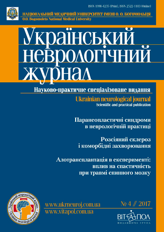
№4(45) // 2017

Paraneoplastic syndromes in neurological practice
E. G. Dubenko, L. I. Kovalenko
Analysis of comorbid diseases in patients with multiple sclerosis
Т. І. Nehrych, К. М. Hychka
Comparative analysis of the rat’s paretic limb spasticity against the background of spinal cord injury, adult olfactory bulb and fetal cerebellum tissue allotransplantation
V. I. Tsymbaliuk 1, 2, V. V. Medvediev 2, Yu. Yu. Senchyk 3, N. G. Draguntsova 1
Log In
Notice: Undefined variable: err in /home/vitapol/ukrneuroj.vitapol.com.ua/blocks/news.php on line 50

