- Issues
- About the Journal
- News
- Cooperation
- Contact Info
Issue. Articles
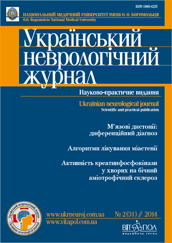
№2(31) // 2014

1.
|
Notice: Undefined index: picture in /home/vitapol/ukrneuroj.vitapol.com.ua/en/svizhij_nomer.php on line 74 Notice: Undefined index: pict in /home/vitapol/ukrneuroj.vitapol.com.ua/en/svizhij_nomer.php on line 75 Muscular dystonia. Analytical review and own clinical observationT. V. Myronenko, O. Yu. Rybalka, M. O. Myronenko, I. V. Hubetova, M. K. Gaidash |
|---|
The authors summarized the literature on the etiology, pathogenesis, classification, clinics and diagnostics muscular dystonia. The differentiation of the individual forms of dystonia is presented. The international clinical diagnostic criteria for this disease are outlined. As an example, own clinical observation of the patient with muscular dystonia is introduced. The above information expands the idea about the features of muscular dystonia late manifestation.
Keywords: muscular dystonia, clinics, diagnostics.
Notice: Undefined variable: lang_long in /home/vitapol/ukrneuroj.vitapol.com.ua/en/svizhij_nomer.php on line 188
2.
|
Notice: Undefined index: picture in /home/vitapol/ukrneuroj.vitapol.com.ua/en/svizhij_nomer.php on line 74 Notice: Undefined index: pict in /home/vitapol/ukrneuroj.vitapol.com.ua/en/svizhij_nomer.php on line 75 Myasthenia gravis: modern approaches to diagnostic and treatmentV. M. Skolnyk, O. I. Kalbus, O. M. Baranenko, O. V. Pogorielov |
|---|
The modern concept of pathogenesis and clinical course of myasthenia gravis are presented. The diagnostic and treatment approaches are emphasized.
Keywords: myasthenia gravis, ocular form, generalized form, diagnostic, treatment.
Notice: Undefined variable: lang_long in /home/vitapol/ukrneuroj.vitapol.com.ua/en/svizhij_nomer.php on line 188
3.
|
Notice: Undefined index: picture in /home/vitapol/ukrneuroj.vitapol.com.ua/en/svizhij_nomer.php on line 74 Notice: Undefined index: pict in /home/vitapol/ukrneuroj.vitapol.com.ua/en/svizhij_nomer.php on line 75 Poststroke spasticity: pathophysiology and methods of diagnostics (review of the problem)А. V. Раyenok, I. M. Tsiumrak, S. Yа. Kyrylyuk |
|---|
The article deals with the latest data of the pathophysiology of poststroke spasticity resarches, methods of study with a detailed general, neurological examination and electroneuromiography. Consistent and correct patient’s examination of poststroke spasticity is important for the determination of the status of the patient, planning effective rehabilitation and dynamic monitoring of treatment in order to reduce the suffering of poststroke spasticity of patients.
Keywords: stroke, spasticity, pathophysiology, methods of study, scales.
Notice: Undefined variable: lang_long in /home/vitapol/ukrneuroj.vitapol.com.ua/en/svizhij_nomer.php on line 188
4.
|
Notice: Undefined index: picture in /home/vitapol/ukrneuroj.vitapol.com.ua/en/svizhij_nomer.php on line 74 Notice: Undefined index: pict in /home/vitapol/ukrneuroj.vitapol.com.ua/en/svizhij_nomer.php on line 75 Connection between clinical characteristics of ischemic stroke and arterial hypertension degreeL. I. Sokolova, T. M. Cherenko, Yu. L. Heletyuk |
|---|
Objective — to investigate the connection between the severity of ischemic stroke and its subtypes, localization and size of ischemia and degree of arterial hypertension.
Methods and subjects. 105 patients with ischemic stroke and arterial hypertension of different degrees (I, II, III) (mean age — 67.30 ± 1.09; females — 50 %; males — 55 %) have been examined. Stroke severity was evaluated by NIHSS, subtypes — by TOAST. The diagnosis of hypertension was based on clinical, laboratory and instrumental data.
Results. The degree of arterial hypertension was significantly correlated with the severity of neurological disorders (r = 0.34). Patients with severe neurological disorders did not have any symptoms of first degree of arterial hypertension. The second degree of arterial hypertension was more spread among patients with ischemic stroke.
Conclusions. The severity of neurological disorders correlated with the degree of arterial hypertension, a significant increase in the proportion of patients with moderately severe and severe neurological deficit with increasing degree of hypertension was found.
Keywords: cerebral ischemic stroke, arterial hypertension, blood pressure.
Notice: Undefined variable: lang_long in /home/vitapol/ukrneuroj.vitapol.com.ua/en/svizhij_nomer.php on line 188
5.
|
Notice: Undefined index: picture in /home/vitapol/ukrneuroj.vitapol.com.ua/en/svizhij_nomer.php on line 74 Notice: Undefined index: pict in /home/vitapol/ukrneuroj.vitapol.com.ua/en/svizhij_nomer.php on line 75 Сreatine kinasemia in amyotrophic lateral sclerosisN. P. Voloshina, O. V. Egorkina, V. V. Ryazantsev, M. E. Chernenko |
|---|
Objective — to evaluate the activity of the enzyme creatine phosphokinase (CPK) of serum and cerebrospinal fluid as a biomarker of the disease in patients with amyotrophic lateral sclerosis (ALS).
Methods and subjects. Patients with ALS were examined. ALS was defined by revised El Escorial diagnostic criteria, electromyographic criteria and measurement criteria of nerve conduction for the diagnosis of ALS. Patients received treatment in the department of neuroinfections and multiple sclerosis of SI INPN NAMSU in 2012 — 2013. CPK activity was determined by a kinetic spectrophotometric method in 52 patients in the serum and in 35 patients in the cerebrospinal fluid. CPK levels in patients with different forms of ALS were considered by gender, ALSFRS-R (Functional Rating Scale ALS, Revised) score and duration of the disease.
Results. Analysis of the finding revealed that the CPK enzyme activity of cerebrospinal fluid of ALS patients was significantly higher than the maximum reference value of CPK activity of cerebrospinal fluid of healthy people; total serum CPK was elevated in 50 % of ALS patients. The mean value of serum CPK level was significantly higher in men than in women; the mean value of serum CPK levels was statistically elevated in men with lumbosacral form of ALS; patients with bulbar form of the disease, regardless of gender, had mean CPK level within the normal range; there was no significant difference in the mean values of serum CPK level of patients with onset of the disease with upper limbs involvement and with lower limbs involvement.
Conclusions. In ALS patients were observed or elevated regulation of CPK. These data of CPK levels of serum and cerebrospinal fluid of ALS patients can be considered as one of the prognostic factors of survival.
Keywords: amyotrophic lateral sclerosis, creatine phosphokinase, survival.
Notice: Undefined variable: lang_long in /home/vitapol/ukrneuroj.vitapol.com.ua/en/svizhij_nomer.php on line 188
6.
|
Notice: Undefined index: picture in /home/vitapol/ukrneuroj.vitapol.com.ua/en/svizhij_nomer.php on line 74 Notice: Undefined index: pict in /home/vitapol/ukrneuroj.vitapol.com.ua/en/svizhij_nomer.php on line 75 Changes in lipid peroxidation, antioxidant system, platelet hemostasis in opiate addicted patients depending on the presence of antibodies to HIV 1/2I. I. Bokal, V. V. Kostyushov, N. V. Klimchenko |
|---|
Objective — to examine changes in the POL/AOS and platelet link of hemostasis in patients with opiate addiction, depending on the presence or absence of antibodies to HIV 1/2.
Methods and subjects. The paper presents the results of 30 patients examination with opiate injection drug aged 24 to 45 years (of them men — 27, women — 3) being treated at the Medical Center Polinar. All respondents were divided into 2 groups. Group 1 — 15 addicts who had antibodies to HIV 1/2 (HIV 1/2) in serum, and 80 % of cases with antibodies for hepatitis C (HCV). Group 2 included 15 HIV-uninfected drug addicted with the antibodies for hepatitis C. Antibodies to hepatitis B surface antigen (HBsAg), at the time of examination, in patients 1 and 2 groups were not detected. The comparison group consisted of 20 patients with antibodies to hepatitis C. The control group consisted of 100 healthy people blood donors. Investigation of processes POL / AOS and platelet hemostasis was performed in serum samples of the groups obtained from venous blood by standard methods before starting therapy.
Results. The clinical picture of drug addiction in both groups was typical for this disease: cytolytic syndrome was observed in all patients and was moderate in HIV-uninfected drug users. All addicted had decrease in the level of glucose to 3.0 mmol/l. Thus, HIV-independent patients had lowered platelet by 56 % compared with the control group. In HIV-infected drug users demonstrated more pronounced thrombocytopenia, platelet count decreased by 68 %. Patients with hepatitis C platelet count decreased by 44 %. Content of total antioxidants decreased by 56 %, 34 % and 14 %, respectively. However, not only in HIV-infected drug users, but also in HIV-uninfected patients (HIV- uninfected drug users, hepatitis C) observed identical in nature of abnormal TDS, which revealed increased content of oxidized forms of proteins and reduced forms of the low molecular weight thiol compounds. These changes were more pronounced in HIV-infected drug users. It evidences the nonspecific nature of the changes noted by TDS. Therefore, TDS can be considered as a system, which is directly involved in the molecular mechanisms of nonspecific resistance to damaging factors.
Conclusions. Lipid peroxidation, antioxidant system processes are activated by the delay of malondialdehyde neutralization and utilization, accumulation of diene conjugate. These disorders are more marked in HIV-infected drug users. Different by origin factors (viral and opiate load) influence can cause unidirectional changes of oxide regeneration balance in TDS which is an important part of nonspecific organism resistance. The study of lipid peroxidation, antioxidant defense system status and their clinical evaluation in drug addicted is not only an important from a scientific point of view, but it has practical value in terms of an adequate therapeutic programs using antioxidants.
Keywords: opiate addiction, lipid peroxidation, antioxidant system, thiol-disulfide system, platelet hemostasis.
Notice: Undefined variable: lang_long in /home/vitapol/ukrneuroj.vitapol.com.ua/en/svizhij_nomer.php on line 188
7.
|
Notice: Undefined index: picture in /home/vitapol/ukrneuroj.vitapol.com.ua/en/svizhij_nomer.php on line 74 Notice: Undefined index: pict in /home/vitapol/ukrneuroj.vitapol.com.ua/en/svizhij_nomer.php on line 75 Epidemiological and clinical features of multiple sclerosis in LutskO. D. Shulga, L. A. Shulga, S. Yu. Boyko, O. S. Drozd, Zh. O. Matvejchuk, S. V. Melnik, C. F. Melnik, O. Ya. Oranska, O. M. Yunchik |
|---|
Objective — to study clinical and epidemiological features in multiple sclerosis (MS) patients in Lutsk.
Methods and subjects. 153 patients with MS citizens of Lutsk city were examined. Data concerning age, gender, onset of the disease, course and disability were analyzed. The prevalence and incidence of MS in Lutsk during last 15 years were analyzed. The MS prevalence in age and gender groups has been observed for over 15 years.
Results. Lutsk city belongs to the high zone risk of multiple sclerosis with prevalence of 113.7 per 100,000. Female to male ratio is 2.3 : 1. There is a slight tendency to decrease of incidence and prevalence of MS for the last 5 years. relapsing-remmiting and relapsing-progressive type of disease are dominant. Primary-progressive MS was observed in 7.19 % of cases. The highest prevalence of MS was observed in the age groups after 50 years. The mean age for disease onset is 34.3 years in male and 37.6 years in female.
Conclusions. High level of disability indicates the necessity of rehabilitation system with MS.
Keywords: multiple sclerosis, incidence, prevalence, type of disease.
Notice: Undefined variable: lang_long in /home/vitapol/ukrneuroj.vitapol.com.ua/en/svizhij_nomer.php on line 188
8.
|
Notice: Undefined index: picture in /home/vitapol/ukrneuroj.vitapol.com.ua/en/svizhij_nomer.php on line 74 Notice: Undefined index: pict in /home/vitapol/ukrneuroj.vitapol.com.ua/en/svizhij_nomer.php on line 75 Peculiarities of vegetative regulation among young men sufferering from cardiac pathologyО. V. Romaliyskaya, А. V. Demchenko, А. V. Revenko, О. V. Nickityuk, V. А. Kharyakov |
|---|
Objective — to study functions of VNS by means of the estimation of different vegetative regulation complexes and using the methods of evoked skin sympathetic potentials (ESSP) among young men suffering from cardiac pathology in comparison with those in good health.
Methods and subjects. 45 young men with the testing in accordance with Vein – Solovyova inquirer, calculation of Kerdo index and Hildebrandt’s coefficient, ESSP investigation with analysis of standard characteristics have been examined. As a result of that a main group (I) contained 30 patients of age limit 20.7 years ± 1.8 with corresponding cardiac pathology. Control group (II) contained 15 healthy people of age limit 20.7 years ± 1.9.
Results. Patients of the I group had total tonus of VNS mainly with sympathetic orientation. Kerdo index in the I group had negative result in 40 % and positive — in 47 %. In the II group it was equal to 0. The calculation of Hildebrandt’s coefficient denotes an error of visceral systems activity in the I group. Latent time of ESSP in the I group was reliably smaller than in the II group (р < 0.05). Аmplitude and duration of the first and the second phases in the I group were reliably higher in comparison with the II group (р < 0.05).
Conclusions. The elevation of ergotropic centers activity was revealed among patients from the I group.
Keywords: vegetative regulation, evoked skin sympathetic potentials, cardial patology.
Notice: Undefined variable: lang_long in /home/vitapol/ukrneuroj.vitapol.com.ua/en/svizhij_nomer.php on line 188
9.
|
Notice: Undefined index: picture in /home/vitapol/ukrneuroj.vitapol.com.ua/en/svizhij_nomer.php on line 74 Notice: Undefined index: pict in /home/vitapol/ukrneuroj.vitapol.com.ua/en/svizhij_nomer.php on line 75 Restorative treatment of patients with а cavernous angioma of the brain stem in early postoperative periodV. M. Zhdanova |
|---|
Objective — to develop the complex of therapeutic measures in the early postoperative period to restore disturbed functions of patients who underwent a surgery for cavernous angioma of the brain stem
Methods and subjects. The article represents the treatment results analysis of 18 patients who underwent a surgery for cavernous angioma of the brain stem. The following methods were applied: physical therapy, medication, physiotherapy.
Results. Restorative treatment started after stabilization of vital functions. Combined physical therapy with potentiating effect were administered: ultrasound therapy, followed by electrical stimulation of facial and eye muscles. In order to restore swallowing electrical stimulation of the pharyngeal muscles was used. All patients had regression of neurological deficit.
Conclusions. The complex of the restorative treatment of patients after a surgery for а cavernous angioma of the brain stem provides restoration of disturbed function, significantly improves the quality of life.
Keywords: cavernous angioma, the early postoperative period, restorative treatment, physical therapy.
Notice: Undefined variable: lang_long in /home/vitapol/ukrneuroj.vitapol.com.ua/en/svizhij_nomer.php on line 188
10.
|
Notice: Undefined index: picture in /home/vitapol/ukrneuroj.vitapol.com.ua/en/svizhij_nomer.php on line 74 Notice: Undefined index: pict in /home/vitapol/ukrneuroj.vitapol.com.ua/en/svizhij_nomer.php on line 75 Brain gliomas: postoperative rehabilitation and patients’ life qualityV. D. Rozumenko, A. P. Khoroshun |
|---|
Objective — to improve life quality of patients with brain gliomas by the introduction of optimal methods of recovery treatment in postoperative period.
Methods and subjects. In accordance with complex clinical and neurological estimation, MRI, CT, MRI, dynamic MRI and SPECT results of 462 patients with brain gliomas the tactics of rehabilitation treatment was developed. Postoperative treatment course included pharmacotherapy, electro-stimulation, laser therapy, medical gymnastic. Postoperative treatment care was arranged taking into account the neurological deficit. localization of a tumor and its position relatively to functionally important zones of the brain, malignancy degree. Comparative analysis of neurological sings of disease in pre- and postoperative periods was performed.
Results. The use of the developed methods of rehabilitation treatment in patients with brain gliomas allows to optimize the treatment of patients in postoperative period. As the results after the surgical removal of a tumor and a rehabilitation treatment the percent of patients with a KPS ³ 70 has increased from 47.3 % to 86.4 %.
Conclusions. The use of described tactics of rehabilitation treatment of patient with brain gliomas provides the ability of social rehabilitation, promotes patients’ life quality.
Keywords: brain gliomas, recovery treatment, quality of life.
Notice: Undefined variable: lang_long in /home/vitapol/ukrneuroj.vitapol.com.ua/en/svizhij_nomer.php on line 188
11.
|
Notice: Undefined index: picture in /home/vitapol/ukrneuroj.vitapol.com.ua/en/svizhij_nomer.php on line 74 Notice: Undefined index: pict in /home/vitapol/ukrneuroj.vitapol.com.ua/en/svizhij_nomer.php on line 75 Intracranial tumors in newbornsYu. O. Orlov, A. V. Shaversky, L. L. Marushchenko, V. I. Zyabchenko |
|---|
Objective — a retrospective study of brain tumors diagnosed during the neonatal period and treated in Pediatric Neurosurgical Department in SI «Institute of Neurosurgery, NAMS of Ukraine» from 1980 till 2012.
Methods and subjects. During 33 years, 15 cases of newborn babies with brain tumors were observed at the Institute of Neurosurgery, which makes 0.3 % of all children cases with brain tumors treated at the Pediatric department over the above mentioned period. Ten of them were boys (66.7 %), and five — girls (33.3 %). The histological analysis of the tumor was confirmed in 13 (86.7 %) observations. Benign tumors were diagnosed in 6 (46.1 %) cases: plexus papilloma — 1, atypical plexus pailloma — 1, hemangiopericytoma — 1, a mature teratoma — 1, and lipomas — 2. Malignant tumors were found in 7 (53.9 %) cases: anaplastic astrocytomas — 3, neuroblastoma — 1, an immature teratoma — 1, medulloblastoma — 1 and sPNET — 1. The histostructure was not identified in 2 cases. CT was performed in 15 cases, MRI — in 14 and neurosonology in 10 cases. Supratentorial tumours were in 13 (86.7 %) patients and subtentorial — in 2 (13.3 %).
Results. The basic clinical symptoms of tumours after birth were: macrocrania, anxiety, pyramid insufficiency, convulsions. The signs of intracranial hypertension were notified in 7 (46.7 %) patients. In total, 11 patients underwent 13 operations, tumor removal was complemented by a shunting procedure in two cases. A total tumor removal was performed in 7 patients, a subtotal tumor removal — in 2 cases and a biopsy — in one case. Two patients died after removing of tumours and 1 — after a shunt operation. The mortality after the tumor removal was 20 %. 7 patients had catamnesis; the average life expectancy was 3.1 years (from 1 to 12 years).
Conclusions. Modern diagnostic techniques make it possible to diagnose the congenital brain tumors at the early stages of life, or prenatally. Aggressive surgical tactic, in relation to most brain tumours in neonatal period, is the most adequate in this situation. Results of research discovered that a basic factor which influenced the prognosis was bleeding during operation and in the late postoperative period the volume of ablated tumour and degree of tumour malignancy. Though the treatment outcomes and the survival rate remain poor, certain categories of children show a quite satisfactory period of outcome and the quality of life.
Keywords: brain neoplasm, neonatal tumors, newborns, neuroepithelial tumors.
Notice: Undefined variable: lang_long in /home/vitapol/ukrneuroj.vitapol.com.ua/en/svizhij_nomer.php on line 188
12.
|
Notice: Undefined index: picture in /home/vitapol/ukrneuroj.vitapol.com.ua/en/svizhij_nomer.php on line 74 Notice: Undefined index: pict in /home/vitapol/ukrneuroj.vitapol.com.ua/en/svizhij_nomer.php on line 75 Clinics and diagnosis of the distal anterior cerebral аrtery aneurysms in acute rupture periodA. D. Sydorak, O. A. Tsimeyko |
|---|
Objective — to determine the typical clinical manifestations of aneurysms of the distal segment of the anterior cerebral artery in acute rupture period, and their diagnostic methods.
Methods and subjects. 54 patients with aneurysms of the distal segment of the anterior cerebral artery in acute rupture period were examined at the Institute of neurosurgery from 1998 to 2013.
Results. The main clinical manifestation of artery aneurisms rupture is subarachnoid hemorrhage. All patients underwent CT of cerebrum, the result of which showed the following data: 15 patients were diagnosed with subarachnoid hemorrhage, 22 patients had subarachnoid parenchymal intracerebral hemorrhage, 10 — subarachnoid parenchymal ventricular hemorrhage, 2 — subarachnoid-ventricular hemorrhage.
Сonclusions. The main diagnostic method for aneurysms of the distal segment of the anterior cerebral artery is cerebral angiography. Highly informative methods are also CT, CT-AG, MRI of the brain.
Keywords: subarachnoid hemorrhage, arterial aneurysm.
Notice: Undefined variable: lang_long in /home/vitapol/ukrneuroj.vitapol.com.ua/en/svizhij_nomer.php on line 188
13.
|
Notice: Undefined index: picture in /home/vitapol/ukrneuroj.vitapol.com.ua/en/svizhij_nomer.php on line 74 Notice: Undefined index: pict in /home/vitapol/ukrneuroj.vitapol.com.ua/en/svizhij_nomer.php on line 75 Wernicke’s encephalopathy: clinical observation of a pregnantV. N. Vasilenko, V. A. Penner, M. O. Myronenko, V. S. Kovalik |
|---|
Wernicke’s encephalopathy — an acute neuropsychiatric syndrome that develops as a result of thiamine deficiency and is accompanied by a significant risk of complications and death. Own clinical observation Wernicke encephalopathy pregnant is described. The need for early diagnosis and timely prevention of the disease is emphasized.
Keywords: Wernicke’s encephalopathy, pregnancy, deficiency of vitamin B1.
Notice: Undefined variable: lang_long in /home/vitapol/ukrneuroj.vitapol.com.ua/en/svizhij_nomer.php on line 188
14.
|
Notice: Undefined index: picture in /home/vitapol/ukrneuroj.vitapol.com.ua/en/svizhij_nomer.php on line 74 Notice: Undefined index: pict in /home/vitapol/ukrneuroj.vitapol.com.ua/en/svizhij_nomer.php on line 75 Problems of diagnosis and treatment of acute porphyric polyneuropathyT. I. Negrych, M. I. Yur, A. I. Khoma, N. M. Kardosh, J. Y. Sanotskyy, N. R. Got |
|---|
The article presents a clinical case of acute porphyric polyneuropathy, the difficulty of timely diagnosis and peculiarities of this disease. Methods for diagnosis of porphyria and drugs for the pathogenetic treatment of disease are absent.
Keywords: heme, porphyria, porphyric polyneuropathy.
Notice: Undefined variable: lang_long in /home/vitapol/ukrneuroj.vitapol.com.ua/en/svizhij_nomer.php on line 188
15.
|
Notice: Undefined index: picture in /home/vitapol/ukrneuroj.vitapol.com.ua/en/svizhij_nomer.php on line 74 Notice: Undefined index: pict in /home/vitapol/ukrneuroj.vitapol.com.ua/en/svizhij_nomer.php on line 75 Systemic tick-borne borreliosis: diagnosis and neuroophthalmological violations therapy optimizationA. A. Rudenko, L. V. Murawska, P. A. Dyachenko, B. A. Parкhomets, V. Y. Lutsenko, J. P. Sidorova |
|---|
Objective — to examine neuroophthalmological disorders in systemic borreliosis.
Methods and subjects. The study involved 30 patients with Lyme borreliosis, including female patients — 18, male patients — 12. 11 patients were 21 — 30 years old, from 31 to 40 years — 6 patients, from 41 to 50 years — 6, from 51 to 60 — 4, 61 years and over — 1. The moderate severity of the disease was observed in 20 patients, severe — 9, mild — 1. The skin lesions in the form of annular erythema were present in 12 patients, musculoskeletal dysfunctions were in 10, the nervous system in the form of disseminated encephalomyelitis were in 2 cases and polyneuropathy was diagnosed in 1case, eye disease was observed in 5 cases, four of them were diagnosed with retrobulbar neuritis, 1patient had chorioretinitis.
Results. All cases of Lyme borreliosis with eye disease were diagnosed late, patients were treated in different hospitals (ocular, neurological) and only after infectiologist’s consultation Lyme borreliosis test was prescribed. It should be noted that cases of eye disease were in non erythemal Lyme borreliosis form. In addition, all patients lived in endemic Lyme borreliosis areas, but only two patients removed ticks, the other three did not remember this. The history case of those two patients made it possible to suspect Lyme borreliosis, a diagnosis that was made after the laboratory tests. Three patients, who did not remove ticks, suffered from neuroophthalmological violations against the background of herpes virus infection in a state of persistence. They were examined for Lyme borreliosis by specific serological tests that confirmed the diagnosis of Lyme borreliosis.
Conclusions. In ophthalmological disorders such as retrobulbar neuritis, neuroritinitis, chorioretinitis, keratitis one should be aware of Lyme borreliosis and conduct relevant tests for blood antibody titers Ig M, Ig G to Borel. The main method of chronic Lyme borreliosis treatment is the administration of antibiotics in high doses for 21 — 28 days, or to conduct therapy with ceftriaxone 4 grams per day during 4 days, 3 day break and go on throughout the month, in chronic forms it should last longer.
Keywords: system tick borreliosis, neyrooftalmological disorder diagnosis, therapy.
Notice: Undefined variable: lang_long in /home/vitapol/ukrneuroj.vitapol.com.ua/en/svizhij_nomer.php on line 188
16.
|
Notice: Undefined index: picture in /home/vitapol/ukrneuroj.vitapol.com.ua/en/svizhij_nomer.php on line 74 Notice: Undefined index: pict in /home/vitapol/ukrneuroj.vitapol.com.ua/en/svizhij_nomer.php on line 75 The mechanism of acute phase of a facet pain syndrome by the example of a thoracic levelV. V. Gongalsky |
|---|
Objective — to detect the exact anatomical structures in intercostal articulations which may be a source of a nociceptive afferentation in a facet pain syndrome of biomechanical origin.
Methods and subjects. The specific vertebra dislocation in a thoracic spinal unit revealed on X-ray photographs on the thoracic level with a facet pain syndrome were reproduced on 42 animals (rabbits). Tissues of flexible thoracic spinal unit, substantia gelatinosa of Rolando and cells of lateral horn of spinal cord gray material have been investigated on appropriate segments of spinal cord in terms of 7, 14, 28, 42, 56, 84 and 168 days.
Results. The most possible source of nociceptive afferentation in facet pain syndrome in its acute phase has been detected. The most reactive from investigated tissue of intercostal articulations has appeared to be a synovial fold. It reacts on facets displacement by marked edema, mechanically waging in joint cavity and blocking of spinal motional unit. The intact synovia fold is presented for comparison. Revealed accompanying reaction of neurons in substance of gelatinoid of Rolando and cells of intermediolateral nucleus of spinal cord gray material were confirmed as nociceptive afferentation and its vegetative component in a biomechanically origin facet pain syndrome.
Conclusions. One of the most important mechanisms of a facet pain syndrome in acute phase is a marked synovial fold edema and it waging in joint cavity due to the result of extra displacement in intercostal articulations. The rapid development of a synovial fold edema is due to the plenty vascularization of a synovial fold. Nociceptive response is supported by receptors fields of inflamed and squeezed synovial fold. The facet syndrome is connected with neural cells reactions in a spinal cord which participate in arising of pain with a marked vegetative component.
Keywords: facet pain syndrome, spinal pain, synovial fold, zygapophysal joints, nociception of spinal motional unit, gelatinoid substance of Rolando.
Notice: Undefined variable: lang_long in /home/vitapol/ukrneuroj.vitapol.com.ua/en/svizhij_nomer.php on line 188
17.
|
Notice: Undefined index: picture in /home/vitapol/ukrneuroj.vitapol.com.ua/en/svizhij_nomer.php on line 74 Notice: Undefined index: pict in /home/vitapol/ukrneuroj.vitapol.com.ua/en/svizhij_nomer.php on line 75 Neurobiological mechanisms of arterial hypertension formation in rats and its correction with help distant implantation embryonic tissues anterior segments of leechT. M. Vorobiova, О. V. Veselovska, A. V. Shlyahova, Т. І. Garmash |
|---|
Objective — investigation of the influence of distant implantation embryonic tissues anterior segments of leech on changes in hemodynamic parameters and emotional reactions in animals with arterial hypertension.
Methods and subjects. Investigations were carried out on 30 mongrel white male rats. After the formation of arterial hypertension, by creating zoo conflict situation, rats distant implantation embryonic tissues anterior segments leeches subcutaneously was conducted in the region of projection of the third cervical vertebra. Measurement of systolic blood pressure, the latent period of immobility, ECG registration were performed in intact animals, after the formation of arterial hypertension and after distant implantation embryonic tissues anterior segments of leeches.
Results. The obtained results of the statistical and correlation analysis of heart rate variability, the latent period of immobility in the test «hanging by the tail» illustrate the active participation of the sympathetic autonomic nervous system in the regulation of cardiovascular activity in rats with arterial hypertension. Increased parasympathetic influences autonomic nervous system was observed after distant implantation of the anterior segments embryonic leech in comparison with indicators of stress after — effects. It should be noted that distant implantation embryonic tissues anterior divisions of leech is minimally invasive in comparison with intracerebral transplantation and has long-lasting effect in comparison with traditional hirudotherapy.
Conclusions. It has been showed a positive effect of distant implantation embryonic tissues anterior segments of leech in rats with arterial hypertension, as evidenced by normalization of systolic blood pressure against the background of activation of adaptive systems of animals.
Keywords: arterial hypertension, distant implantation embryonic tissues anterior divisions of leeches.
Notice: Undefined variable: lang_long in /home/vitapol/ukrneuroj.vitapol.com.ua/en/svizhij_nomer.php on line 188
18.
|
Notice: Undefined index: picture in /home/vitapol/ukrneuroj.vitapol.com.ua/en/svizhij_nomer.php on line 74 Notice: Undefined index: pict in /home/vitapol/ukrneuroj.vitapol.com.ua/en/svizhij_nomer.php on line 75 Мышечные дистонии. Аналитический обзор и собственное клиническое наблюдениеТ. В. Мироненко, О. Ю. Рыбалка, М. О. Мироненко, И. В. Хубетова, М. К. Гайдаш |
|---|
Обобщены данные литературы по вопросам этиологии, патогенеза, классификации, клинических проявлений и диагностики мышечных дистоний. Проведена дифференциация отдельных форм дистоний. Освещены международные клинико-диагностические критерии данного заболевания. Приведено собственное клиническое наблюдение пациентки с миодистонией. Изложенная информация расширяет представление об особенностях поздней манифестации мышечной дистонии.
Keywords: мышечная дистония, клинические проявления, диагностика.
Notice: Undefined variable: lang_long in /home/vitapol/ukrneuroj.vitapol.com.ua/en/svizhij_nomer.php on line 188
19.
|
Notice: Undefined index: picture in /home/vitapol/ukrneuroj.vitapol.com.ua/en/svizhij_nomer.php on line 74 Notice: Undefined index: pict in /home/vitapol/ukrneuroj.vitapol.com.ua/en/svizhij_nomer.php on line 75 Миастения: современные подходы к диагностике и лечениюВ. М. Школьник, А. И. Кальбус, А. Н. Бараненко, А. В. Погорелов |
|---|
Приведены современные представления о патогенезе и клиническом течении миастении. Особое внимание уделено диагностическим и лечебным подходам.
Keywords: миастения, глазная форма, генерализованная форма, диагностика, лечение.
Notice: Undefined variable: lang_long in /home/vitapol/ukrneuroj.vitapol.com.ua/en/svizhij_nomer.php on line 188
20.
|
Notice: Undefined index: picture in /home/vitapol/ukrneuroj.vitapol.com.ua/en/svizhij_nomer.php on line 74 Notice: Undefined index: pict in /home/vitapol/ukrneuroj.vitapol.com.ua/en/svizhij_nomer.php on line 75 Креатинкиназемия при боковом амиотрофическом склерозеН. П. Волошина, О. В. Егоркина, В. В. Рязанцев, М. Е. Черненко |
|---|
Цель — оценить активность креатинфосфокиназы (КФК) в сыворотке крови и ликворе больных боковым амиотрофическим склерозом (БАС) в качестве биомаркера заболевания.
Материалы и методы. В исследование были включены больные с диагнозом БАС, установленным на основании пересмотренных диагностических критериев El Escorial, электромиографических критериев и критериев измерения проводимости нервов для диагностики БАС, находившиеся на стационарном лечении в отделе нейроинфекций и рассеянного склероза Института неврологии, психиатрии и наркологии НАМН Украины в 2012 — 2013 гг. Активность КФК определяли кинетическим спектрофотометрическим методом у 52 больных в сыворотке крови и у 35 больных в ликворе. Уровень активности КФК у больных с разными формами БАС изучали с учетом пола, оценки по шкале ALSFRS-R (Functional Rating Scale ALS, Revised) и длительности заболевания.
Результаты. Зафиксировано достоверное повышение активности КФК в ликворе больных БАС по сравнению с показателем пациентов с другими неврологическими заболеваниями и общей КФК в сыворотке крови — у половины больных БАС по сравнению с максимальным референсным значением. Среднее значение уровня КФК в сыворотке крови было достоверно выше у мужчин, в частности у мужчин с пояснично-крестцовой формой БАС. У больных с бульбарной формой заболевания независимо от пола среднее значение содержания КФК было в пределах нормы. Не выявлено достоверных различий уровня КФК в сыворотке крови между больными с началом заболевания с вовлечением верхних или нижних конечностей.
Выводы. У больных БАС отмечена повышенная регуляция КФК. Уровень активности КФК в сыворотке крови и ликворе больных БАС можно рассматривать как один из прогностических факторов выживаемости.
Keywords: боковой амиотрофический склероз, креатинфосфокиназа, выживаемость.
Notice: Undefined variable: lang_long in /home/vitapol/ukrneuroj.vitapol.com.ua/en/svizhij_nomer.php on line 188
Current Issue Highlights
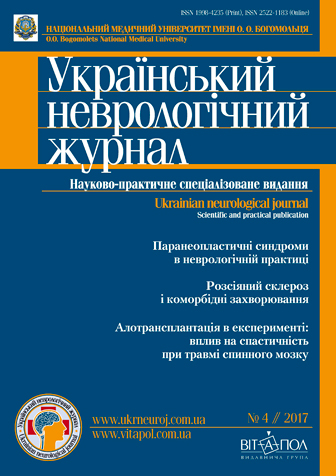
№4(45) // 2017

Paraneoplastic syndromes in neurological practice
E. G. Dubenko, L. I. Kovalenko
Analysis of comorbid diseases in patients with multiple sclerosis
Т. І. Nehrych, К. М. Hychka
Comparative analysis of the rat’s paretic limb spasticity against the background of spinal cord injury, adult olfactory bulb and fetal cerebellum tissue allotransplantation
V. I. Tsymbaliuk 1, 2, V. V. Medvediev 2, Yu. Yu. Senchyk 3, N. G. Draguntsova 1
Log In
Notice: Undefined variable: err in /home/vitapol/ukrneuroj.vitapol.com.ua/blocks/news.php on line 50

